
Pain Physiology – Four images
€ 22,15 Inc. VAT
Description
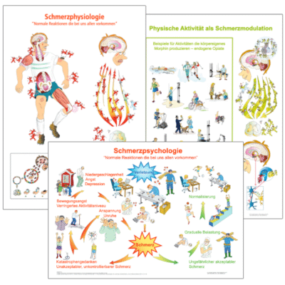
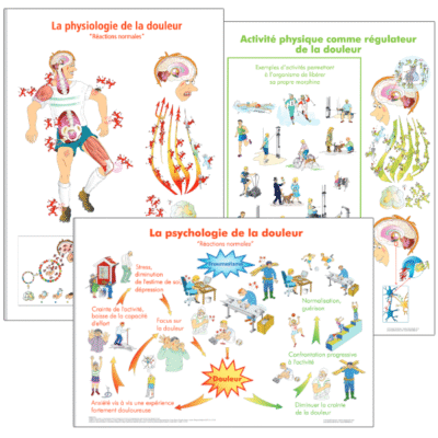
Frame the drawings (A3 format) and put them on the wall in your clinic. Preferably in an area where there is not much going on. In a quiet area, where the patient can look at the drawings not being disturbed. Now the person can observe the drawing and reflect on what the drawing is saying. The person is mirroring her/himself in the drawing, maybe recognizing him/herself. When the patient start to talk about how he/she see, interpreting the information from the drawing, you can add information to what the person is telling you. It is better to guide the patient instead of explaining. This is a biopsychosocial form of communication avoiding the problem of stigmatization. When you purchase these drawings (A3 format) you will also receive a pdf with an explanatory text that you can print and give to your patient. In this way you can use the drawings as a tool combined with other methods/techniques you use helping patients in pain.
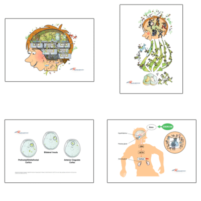
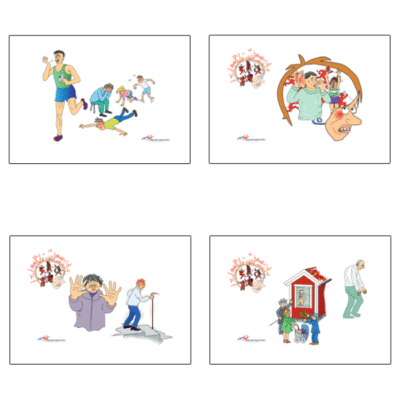
Picture 1 – We have NO pain receptors we have nociceptive receptors

Stroke the skin of your arm carefully. You feel that you stroke your arm. You have now activated low treshold mechanoreceptors in the skin. Now push the thumb hard into the arm. Keep the pressure. After a while it becomes uncomfortable, you experience pain. You may now think that you have activated pain receptors in the tissue. That is not correct. You have activated high treshold nociceptors. Initially, to activate nociceptors (mechanical, thermal and chemical), the stimulus has to be at a certain intensity.
Picture 2 – Nerve impulses from nociceptive receptors are on their way to the brain
 When you suffer an acute tissue injury, high treshold nocicpetors are depolarized. Their nerve impulses travel very fast to the brain making it possible for you to act, avoiding further injury. Nerveimpulses can also travel slower, in so called C fibers. When the nerveimpulses from the nociceptors reach different parts of the brain, primarily insula, and are interpreted as a danger, you experience pain. Thus pain is an output, a feeling, activating a neuromatrix in your brain based on earlier similar experiences. If the pain continue to bother you, the orchestra in your brain start to play pain tunes.
Picture 3 – The negative effects of pain affect whole body systems
 When the orchestra in the brain continue to play pain tunes it will affecting whole body systems where your endocrine-, and immune system will become imbalanced. The chemist shop in your brain stop producing important pain modualting hormones and neurotrasmittors like endorphins, serotonine, oxytocin and cortisol. Because the nervous system is plastic, this situation will make you more prone to negative sensitizations processes resulting in allodynia and hyperalgesia. Ask your doctor/therapist to give you clinical examples of allodynia and hyperalgesia
Picture 4 – Pain can be very stressful
 Over time and due to long term pain, the endocrine system´s HPA axis becomes exhausted resulting in a decrease, or underproduction of cortisol making you more prone to sensitization processes and systematic diseases. Making you more prone to allodyni, which is a pain experience on a normal stimulus that should not cause pain
Related products
-
Exercise for the modulation of pain – Four pictures – digital download
€ 18,00 Inc. VAT Add to cart -
Deutsch – Drei Poster des Schmerzes 50x70cm
€ 59,82 Inc. VAT Add to cart -
Psychology of pain – Four pictures- digital download
€ 18,00 Inc. VAT Add to cart -
French – Trois affiches de douleur – 50x70cm
€ 59,82 Inc. VAT Add to cart